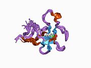
Plasmin
Enzyme in human blood that degrades clots and other proteins
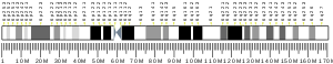
| PLG | |||||||||||||||||||||||||||||||||||||||||||||||||||
|---|---|---|---|---|---|---|---|---|---|---|---|---|---|---|---|---|---|---|---|---|---|---|---|---|---|---|---|---|---|---|---|---|---|---|---|---|---|---|---|---|---|---|---|---|---|---|---|---|---|---|---|
 | |||||||||||||||||||||||||||||||||||||||||||||||||||
| Identifiers | |||||||||||||||||||||||||||||||||||||||||||||||||||
| Aliases | PLG, plasminogen, plasmin, HAE4 | ||||||||||||||||||||||||||||||||||||||||||||||||||
| External IDs | OMIM: 173350; MGI: 97620; HomoloGene: 55452; GeneCards: PLG; OMA:PLG - orthologs | ||||||||||||||||||||||||||||||||||||||||||||||||||
| |||||||||||||||||||||||||||||||||||||||||||||||||||
| |||||||||||||||||||||||||||||||||||||||||||||||||||
| |||||||||||||||||||||||||||||||||||||||||||||||||||
| |||||||||||||||||||||||||||||||||||||||||||||||||||
| |||||||||||||||||||||||||||||||||||||||||||||||||||
| Wikidata | |||||||||||||||||||||||||||||||||||||||||||||||||||
| |||||||||||||||||||||||||||||||||||||||||||||||||||


Plasmin is an important enzyme (EC 3.4.21.7) present in blood that degrades many blood plasma proteins, including fibrin clots. The degradation of fibrin is termed fibrinolysis. In humans, the plasmin protein (in the zymogen form of plasminogen) is encoded by the PLG gene.[5]
Function

Plasmin is a serine protease that acts to dissolve fibrin blood clots. Apart from fibrinolysis, plasmin proteolyses proteins in various other systems: It activates collagenases, some mediators of the complement system, and weakens the wall of the Graafian follicle, leading to ovulation. Plasmin is also integrally involved in inflammation.[6] It cleaves fibrin, fibronectin, thrombospondin, laminin, and von Willebrand factor. Plasmin, like trypsin, belongs to the family of serine proteases.
Plasmin is released as a zymogen called plasminogen (PLG) from the liver into the systemic circulation. Two major glycoforms of plasminogen are present in humans - type I plasminogen contains two glycosylation moieties (N-linked to N289 and O-linked to T346), whereas type II plasminogen contains only a single O-linked sugar (O-linked to T346). Type II plasminogen is preferentially recruited to the cell surface over the type I glycoform. Conversely, type I plasminogen appears more readily recruited to blood clots.
In circulation, plasminogen adopts a closed, activation-resistant conformation. Upon binding to clots, or to the cell surface, plasminogen adopts an open form that can be converted into active plasmin by a variety of enzymes, including tissue plasminogen activator (tPA), urokinase plasminogen activator (uPA), kallikrein, and factor XII (Hageman factor). Fibrin is a cofactor for plasminogen activation by tissue plasminogen activator. Urokinase plasminogen activator receptor (uPAR) is a cofactor for plasminogen activation by urokinase plasminogen activator. The conversion of plasminogen to plasmin involves the cleavage of the peptide bond between Arg-561 and Val-562.[5][7][8][9]
Plasmin cleavage produces angiostatin.
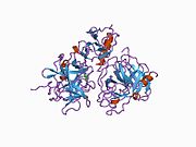
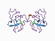
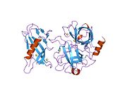
Mechanism of plasminogen activation
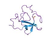
Full length plasminogen comprises seven domains. In addition to a C-terminal chymotrypsin-like serine protease domain, plasminogen contains an N-terminal Pan Apple domain (PAp) together with five Kringle domains (KR1-5). The Pan-Apple domain contains important determinants for maintaining plasminogen in the closed form, and the kringle domains are responsible for binding to lysine residues present in receptors and substrates.
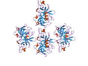
The X-ray crystal structure of closed plasminogen reveals that the PAp and SP domains maintain the closed conformation through interactions made throughout the kringle array .[9] Chloride ions further bridge the PAp / KR4 and SP / KR2 interfaces, explaining the physiological role of serum chloride in stabilizing the closed conformer. The structural studies also reveal that differences in glycosylation alter the position of KR3. These data help explain the functional differences between the type I and type II plasminogen glycoforms.[citation needed]
In closed plasminogen, access to the activation bond (R561/V562) targeted for cleavage by tPA and uPA is blocked through the position of the KR3/KR4 linker sequence and the O-linked sugar on T346. The position of KR3 may also hinder access to the activation loop. The Inter-domain interactions also block all kringle ligand-binding sites apart from that of KR-1, suggesting that the latter domain governs pro-enzyme recruitment to targets. Analysis of an intermediate plasminogen structure suggests that plasminogen conformational change to the open form is initiated through KR-5 transiently peeling away from the PAp domain. These movements expose the KR5 lysine-binding site to potential binding partners, and suggest a requirement for spatially distinct lysine residues in eliciting plasminogen recruitment and conformational change respectively.[9]
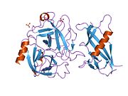
Mechanism of plasmin inactivation
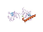
Plasmin is inactivated by proteins such as α2-macroglobulin and α2-antiplasmin.[10] The mechanism of plasmin inactivation involves the cleavage of an α2-macroglobulin at the bait region (a segment of the aM that is particularly susceptible to proteolytic cleavage) by plasmin. This initiates a conformational change such that the α2-macroglobulin collapses about the plasmin. In the resulting α2-macroglobulin-plasmin complex, the active site of plasmin is sterically shielded, thus substantially decreasing the plasmin's access to protein substrates. Two additional events occur as a consequence of bait region cleavage, namely (i) a h-cysteinyl-g-glutamyl thiol ester of the α2-macroglobulin becomes highly reactive and (ii) a major conformational change exposes a conserved COOH-terminal receptor binding domain. The exposure of this receptor binding domain allows the α2-macroglobulin protease complex to bind to clearance receptors and be removed from circulation.
Pathology
Plasmin deficiency may lead to thrombosis, as the clots are not adequately degraded. Plasminogen deficiency in mice leads to defective liver repair,[11] defective wound healing, reproductive abnormalities.[12] [13]
In humans, a rare disorder called plasminogen deficiency type I (Online Mendelian Inheritance in Man (OMIM): 217090) is caused by mutations of the PLG gene and is often manifested by ligneous conjunctivitis.[14]
A rare missense mutation within the kringle 3 domain of plasminogen, resulting in a novel type of dysplasminogenemia, represents the molecular basis of a subtype of hereditary angioedema with normal C1-inhibitor;[15] the mutation creates a new lysine-binding site within kringle 3 and alters the glycosylation of plasminogen.[15] The mutant plasminogen protein has been shown to be a highly efficient kininogenase that directly releases bradykinin from high- and low-molecular-weight kininogen.[16]
Interactions
Plasmin has been shown to interact with Thrombospondin 1,[17][18] Alpha 2-antiplasmin[19][20] and IGFBP3.[21] Moreover, plasmin induces the generation of bradykinin in mice and humans through high-molecular-weight kininogen cleavage.[22]
References
- ^ a b c GRCh38: Ensembl release 89: ENSG00000122194 – Ensembl, May 2017
- ^ a b c GRCm38: Ensembl release 89: ENSMUSG00000059481 – Ensembl, May 2017
- ^ "Human PubMed Reference:". National Center for Biotechnology Information, U.S. National Library of Medicine.
- ^ "Mouse PubMed Reference:". National Center for Biotechnology Information, U.S. National Library of Medicine.
- ^ a b "Entrez Gene: plasminogen".
- ^ Atsev S, Tomov N (December 2020). "Using antifibrinolytics to tackle neuroinflammation". Neural Regeneration Research. 15 (12): 2203–2206. doi:10.4103/1673-5374.284979. PMC 7749481. PMID 32594031.
- ^ Miyata T, Iwanaga S, Sakata Y, Aoki N (October 1982). "Plasminogen Tochigi: inactive plasmin resulting from replacement of alanine-600 by threonine in the active site". Proceedings of the National Academy of Sciences of the United States of America. 79 (20): 6132–6136. Bibcode:1982PNAS...79.6132M. doi:10.1073/pnas.79.20.6132. PMC 347073. PMID 6216475.
- ^ Forsgren M, Råden B, Israelsson M, Larsson K, Hedén LO (March 1987). "Molecular cloning and characterization of a full-length cDNA clone for human plasminogen". FEBS Letters. 213 (2): 254–260. doi:10.1016/0014-5793(87)81501-6. PMID 3030813. S2CID 9075872.
- ^ a b c Law RH, Caradoc-Davies T, Cowieson N, Horvath AJ, Quek AJ, Encarnacao JA, et al. (March 2012). "The X-ray crystal structure of full-length human plasminogen". Cell Reports. 1 (3): 185–190. doi:10.1016/j.celrep.2012.02.012. PMID 22832192.
- ^ Wu G, Quek AJ, Caradoc-Davies TT, Ekkel SM, Mazzitelli B, Whisstock JC, Law RH (April 2019). "Structural studies of plasmin inhibition". Biochemical Society Transactions. 47 (2): 541–557. doi:10.1042/bst20180211. PMID 30837322. S2CID 73463150.
- ^ Bezerra JA, Bugge TH, Melin-Aldana H, Sabla G, Kombrinck KW, Witte DP, Degen JL (December 1999). "Plasminogen deficiency leads to impaired remodeling after a toxic injury to the liver". Proceedings of the National Academy of Sciences of the United States of America. 96 (26): 15143–15148. Bibcode:1999PNAS...9615143B. doi:10.1073/pnas.96.26.15143. PMC 24787. PMID 10611352.
- ^ Romer J, Bugge TH, Pyke C, Lund LR, Flick MJ, Degen JL, Dano K (March 1996). "Impaired wound healing in mice with a disrupted plasminogen gene". Nature Medicine. 2 (3): 287–292. doi:10.1038/nm0396-287. PMID 8612226. S2CID 29981847.
- ^ Ploplis VA, Carmeliet P, Vazirzadeh S, Van Vlaenderen I, Moons L, Plow EF, Collen D (November 1995). "Effects of disruption of the plasminogen gene on thrombosis, growth, and health in mice". Circulation. 92 (9): 2585–2593. doi:10.1161/01.cir.92.9.2585. PMID 7586361.
- ^ Schuster V, Hügle B, Tefs K (December 2007). "Plasminogen deficiency". Journal of Thrombosis and Haemostasis. 5 (12): 2315–2322. doi:10.1111/j.1538-7836.2007.02776.x. PMID 17900274.
- ^ a b Dewald G (March 2018). "A missense mutation in the plasminogen gene, within the plasminogen kringle 3 domain, in hereditary angioedema with normal C1 inhibitor". Biochemical and Biophysical Research Communications. 498 (1): 193–198. doi:10.1016/j.bbrc.2017.12.060. PMID 29548426.
- ^ Dickeson SK, Kumar S, Sun MF, Mohammed BM, Phillips DR, Whisstock JC, et al. (May 2022). "A mechanism for hereditary angioedema caused by a lysine 311-to-glutamic acid substitution in plasminogen". Blood. 139 (18): 2816–2829. doi:10.1182/blood.2021012945. PMC 9074402. PMID 35100351.
- ^ Silverstein RL, Leung LL, Harpel PC, Nachman RL (November 1984). "Complex formation of platelet thrombospondin with plasminogen. Modulation of activation by tissue activator". The Journal of Clinical Investigation. 74 (5): 1625–1633. doi:10.1172/JCI111578. PMC 425339. PMID 6438154.
- ^ DePoli P, Bacon-Baguley T, Kendra-Franczak S, Cederholm MT, Walz DA (March 1989). "Thrombospondin interaction with plasminogen. Evidence for binding to a specific region of the kringle structure of plasminogen". Blood. 73 (4): 976–982. doi:10.1182/blood.V73.4.976.976. PMID 2522013.
- ^ Wiman B, Collen D (September 1979). "On the mechanism of the reaction between human alpha 2-antiplasmin and plasmin". The Journal of Biological Chemistry. 254 (18): 9291–9297. doi:10.1016/S0021-9258(19)86843-6. PMID 158022.
- ^ Shieh BH, Travis J (May 1987). "The reactive site of human alpha 2-antiplasmin". The Journal of Biological Chemistry. 262 (13): 6055–6059. doi:10.1016/S0021-9258(18)45536-6. PMID 2437112.
- ^ Campbell PG, Durham SK, Suwanichkul A, Hayes JD, Powell DR (August 1998). "Plasminogen binds the heparin-binding domain of insulin-like growth factor-binding protein-3". The American Journal of Physiology. 275 (2): E321–E331. doi:10.1152/ajpendo.1998.275.2.E321. PMID 9688635.
- ^ Marcos-Contreras OA, Martinez de Lizarrondo S, Bardou I, Orset C, Pruvost M, Anfray A, et al. (November 2016). "Hyperfibrinolysis increases blood-brain barrier permeability by a plasmin- and bradykinin-dependent mechanism". Blood. 128 (20): 2423–2434. doi:10.1182/blood-2016-03-705384. PMID 27531677.
Further reading
- Shanmukhappa K, Mourya R, Sabla GE, Degen JL, Bezerra JA (July 2005). "Hepatic to pancreatic switch defines a role for hemostatic factors in cellular plasticity in mice". Proceedings of the National Academy of Sciences of the United States of America. 102 (29): 10182–10187. Bibcode:2005PNAS..10210182S. doi:10.1073/pnas.0501691102. PMC 1177369. PMID 16006527.
- Anglés-Cano E, Rojas G (January 2002). "Apolipoprotein(a): structure-function relationship at the lysine-binding site and plasminogen activator cleavage site". Biological Chemistry. 383 (1): 93–99. doi:10.1515/BC.2002.009. PMID 11928826. S2CID 29248198.
- Ranson M, Andronicos NM (May 2003). "Plasminogen binding and cancer: promises and pitfalls". Frontiers in Bioscience. 8 (6): s294–s304. doi:10.2741/1044. PMID 12700073.
External links
- The MEROPS online database for peptidases and their inhibitors: S01.233 Archived 2019-09-13 at the Wayback Machine
- Plasmin at the U.S. National Library of Medicine Medical Subject Headings (MeSH)
This article incorporates text from the United States National Library of Medicine, which is in the public domain.
- v
- t
- e
PDB gallery
-
 1b2i: KRINGLE 2 DOMAIN OF HUMAN PLASMINOGEN: NMR SOLUTION STRUCTURE OF TRANS-4-AMINOMETHYLCYCLOHEXANE-1-CARBOXYLIC ACID (AMCHA) COMPLEX
 -
 1bml: COMPLEX OF THE CATALYTIC DOMAIN OF HUMAN PLASMIN AND STREPTOKINASE
 -
 1bui: STRUCTURE OF THE TERNARY MICROPLASMIN-STAPHYLOKINASE-MICROPLASMIN COMPLEX: A PROTEINASE-COFACTOR-SUBSTRATE COMPLEX IN ACTION.
 -
 1cea: THE STRUCTURE OF THE NON-COVALENT COMPLEX OF RECOMBINANT KRINGLE 1 DOMAIN OF HUMAN PLASMINOGEN WITH EACA (EPSILON-AMINOCAPROIC ACID)
 -
 1ceb: THE STRUCTURE OF THE NON-COVALENT COMPLEX OF RECOMBINANT KRINGLE 1 DOMAIN OF HUMAN PLASMINOGEN WITH AMCHA (TRANS-4-AMINOMETHYLCYCLOHEXANE-1-CARBOXYLIC ACID)
 -
 1ddj: CRYSTAL STRUCTURE OF HUMAN PLASMINOGEN CATALYTIC DOMAIN
 -
 1hpj: SOLUTION NMR STRUCTURE OF THE HUMAN PLASMINOGEN KRINGLE 1 DOMAIN COMPLEXED WITH 6-AMINOHEXANOIC ACID AT PH 5.3, 310K, DERIVED FROM RANDOMLY GENERATED STRUCTURES USING SIMULATED ANNEALING, 12 STRUCTURES
 -
 1hpk: SOLUTION NMR STRUCTURE OF THE HUMAN PLASMINOGEN KRINGLE 1 DOMAIN COMPLEXED WITH 6-AMINOHEXANOIC ACID AT PH 5.3, 310K, DERIVED FROM RANDOMLY GENERATED STRUCTURES USING SIMULATED ANNEALING, MINIMIZED AVERAGE STRUCTURE
 -
 1i5k: STRUCTURE AND BINDING DETERMINANTS OF THE RECOMBINANT KRINGLE-2 DOMAIN OF HUMAN PLASMINOGEN TO AN INTERNAL PEPTIDE FROM A GROUP A STREPTOCOCCAL SURFACE PROTEIN
 -
 1ki0: The X-ray Structure of Human Angiostatin
 -
 1krn: STRUCTURE OF KRINGLE 4 AT 4C TEMPERATURE AND 1.67 ANGSTROMS RESOLUTION
 -
 1l4d: CRYSTAL STRUCTURE OF MICROPLASMINOGEN-STREPTOKINASE ALPHA DOMAIN COMPLEX
 -
 1l4z: X-RAY CRYSTAL STRUCTURE OF THE COMPLEX OF MICROPLASMINOGEN WITH ALPHA DOMAIN OF STREPTOKINASE IN THE PRESENCE CADMIUM IONS
 -
 1pk4: CRYSTAL AND MOLECULAR STRUCTURE OF HUMAN PLASMINOGEN KRINGLE 4 REFINED AT 1.9-ANGSTROMS RESOLUTION
 -
 1pkr: THE STRUCTURE OF RECOMBINANT PLASMINOGEN KRINGLE 1 AND THE FIBRIN BINDING SITE
 -
 1pmk: KRINGLE-KRINGLE INTERACTIONS IN MULTIMER KRINGLE STRUCTURES
 -
 1qrz: CATALYTIC DOMAIN OF PLASMINOGEN
 -
 1rjx: Human PLASMINOGEN CATALYTIC DOMAIN, K698M MUTANT
 -
 2doh: The X-ray crystallographic structure of the angiogenesis inhibitor, angiostatin, bound a to a peptide from the group A streptococcal surface protein PAM
 -
 2doi: The X-ray crystallographic structure of the angiogenesis inhibitor, angiostatin, bound to a peptide from the group A streptococcus protein PAM
 -
 2pk4: THE REFINED STRUCTURE OF THE EPSILON-AMINOCAPROIC ACID COMPLEX OF HUMAN PLASMINOGEN KRINGLE
 -
 5hpg: STRUCTURE AND LIGAND DETERMINANTS OF THE RECOMBINANT KRINGLE 5 DOMAIN OF HUMAN PLASMINOGEN


Portal:
 Biology


